Small devices
MEG lab accelerometers
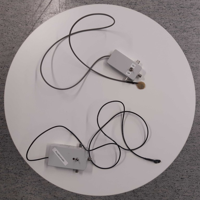
MEB lab has two accelerometers. Both of the accelerometers consist of an accelerometer chip connected with a cable to a box with three BNC connectors. Both of the models have an on/off switch, but only the other one has an “on” indicator LED.
The accelerometers work with 2x1.5V batteries. The LED indicator has one separate 1.5V battery for its power source.
The BNC connectors give out max 3V signal and they represent the X, Y and Z side movement of the accelerometer chip. Normally, the X, Y and Z signals are taken to MEG MISC7-9 channels situated next to the Data Acquisition computer. One should always measure from all the 3 channels which are then quadratically summed in post process to get more real acceleration signal. MISC channels are recorded by turning them on in Acquisition.


MEG lab GSR
MEG lab has one GSR or EDA (skin conductance meter) which is compatible with the Triux MEG. The device consists of Becker Meditec BP-BM-30 (supplier Brain Products) GSR module and Wave-Power-15600 5V USB battery. The GSR is connected to the MEG Bio inputs on the side of the gantry. Because the maximum voltage on these inputs is of the order of +/-15mV, the output voltage of the GSR has to be reduced. This is done with resistors. The output voltage reduction with the current resistor configuration is 200-fold.

If there is no signal reduction, in the output of the GSR module 25 mV corresponds to 1μS with a constant 5V supply voltage. Now, if the USB battery is full, the resistors are exactly what the markings claim them to be, 25mV/200 = 0.125mV corresponds to 1μS.
The participant can be connected to the GSR module with using Ambu single-use skin electrodes. The electrodes can be glued (sticker) to the hand of the participant for example like in the image below. The hand should be the non-dominant hand.

The GSR module is connected to the bioamplifier (BIO) inputs on the side of the MEG gantry (like EOG or ECG cables). Cable marked red to plus, the other to minus.
The USB battery will not stay on automatically by just plugging the GSR module in to it. Because the required current is so small, the battery will switch off automatically after fer seconds. This is circumvented so that when the measurement is done, there is a small lamp on the USB battery which should be on during the measurement. The lamp is turned on by pressing the only button there is for a while.
UPDATE April 11th 2019: With a new black box this can be circumvented. The box has a large enough resistor inside so that the battery stays on without turning the light on. However, the black box seems to sometimes give some funny noise or glitches. So check that it works properly first, if you want to use it.
The output signal of the GSR module is of slowly changing, DC voltage. In the data acquisition computer the channel settings should be set to
- Amplification: 640 (or 2000 if the signal is small)
- DC input
Because the GSR data is only slowly changing, it is difficult to monitor it on the data acquisition computer. The time scale for the GSR signal change is in the order of seconds and more. The maximum time window which can be monitored in the Dacq computer is only few seconds. Additionally, the signal level can change quite a lot during the measurement. To circumvent these problems, it is possible to monitor the GSR signal using the Linux computer and Fieldtrip buffering of the data on the Dacq computer. To do this, see the instructions below.
Breathing sensors
1) There is one piezo-based breathing sensor by "Spes Medica" in the MEG lab. It has a sensor part (short rubber band which has a green fabric strip connectd) and a Velcro strap. There are two different lengths of Velcro straps depending on the girth of your participant. You can strap the device around your participant either around stomach or chest area. As long as the girth changes depending on the breathing. You can ask the participant to exhale and only then tighten the strap. It has to be comfortably tight, so that the strap lengthens some during breathing, but not overly tight.
The device has two lead wires. Connect those directly to the MEG BIO input, which you have prepared from the MEG Data Acquisition program. The voltage generated by the breathing sensor is so low, that it is perfectly safe to directly connect it to the BIO inputs. When you prepare the BIO channel, set the high pass filter of the channel to DC-mode, because the signal will be only slowly changing. This means, there is no high pass filtering. The amplification can be left to the default value.
Note that the breathing sensor is based on piezo crystal. This means, that the device generates voltage when work is done to the crystal (band lengthens or shortens). When no work is done, the voltage level goes back toward zero line. If the participant is holding his/her breath, you might not be able to differentiate normal exhale or inhale from it in every situation. For normal breathing rhythm, the device should be okay.
2) There is now (April 2020) a new breathing sensor which is based on strain gauges. With this one can more easily monitor breathing when for example the participant is holding his/her breath. The signal will not drop by itself. The device has an aluminum part with strain gauges connected (BEWARE NOT TO OVERBEND THIS), a rubber strap and a grey box with connections, small amount of electronics and a power supply (batteries). The white wires with red- and black-colored connectors go to the BIO-input channels of the MEG gantry. Red to positive and black to negative (MAKE SURE THEY ARE CONNECTED CORRECTLY, small danger of braking the amplifiers of the channel otherwise).

EEG-cap wireless impedance checker
Parts:
- Windows-based touch screen computer
- Wireless USB-antenna
- Impedance checker
The Windows touch screen computer has a separate pdf-file with fairly clear instructions. Due to legal reasons, it will not to be distributed on-line in CIBR web pages. The pdf can be easily found in the Windows touch screen computer.


Using Linux stim computer to read signal from DACQ
These instructions might get handy, if you are using GSR, possibly breathing sensor or you just want to show one of the MEG signals in the shielded room on the projector screen.
- Turn on Linux stimulus computer
- Password is the same as in the other stimulus computer(s)
- Press “Data” to mount the disc drive system properly
- If you want to also show the signal in the projector screen, first turn the screen selector for Linux computer (mechanical device below Windows stimulus computer screen), you might need the projector on also, and then run prepare_screen_for_projector.sh shell script which fix monitor settings properly
- In the data acquisition computer start a terminal window
- Type in “neuromag2ft –bufport 49150” and press enter
- Start the data acquisition software (the software can be on already before, but the fieldtrip script will start running only after you have pressed “Start”)
- In the Linux stimulus computer run gsr-monitor.sh shell script
- Write in the “channel name” box your channel in the exact form as it would appear in the DACQ monitoring screen (for example MEG0123, BIO012, etc).
- Tick on the “Automatic scaling” unless you know what scales to write to Y scales.
- Press “Start monitoring”
- Note that the monitoring might not start immediately but the graph can only flash quickly on and then off. There has to be enough lines of data in the DACQ computer fieldtrip buffer, before the stimulus computer’s program can handle it. The buffer has to be maybe 10 or 20 seconds long first.
- Note also that the monitoring will stop automatically on the stimulus computer at some point. Now, there is some kind of maximum time for monitoring set in the program. This might or might not get fixed at some point.
- More notes: The sending of the signal might stop abruptly. Reason might be that the fieldtrip buffering has crashed. Check if the field trip buffering looks normal. If yes, try first stopping and restarting the signal monitoring. If this does not work, stop the field trip monitoring (Ctrl+C in the terminal window should stop it) and restart. If these do not work, call for help unless you know what you are doing.
- Decimal separator is a dot in the Linux monitoring program.
Audiometer
The Department of Psychology has one common audiometer, model SA-51 from Mediroll Medico-technical Ltd. It can play sounds in the range of 250 Hz to 8000 Hz. Maximum sound level slightly depends on the sound frequency, but is in the range of 80 to 100 dB. The device consists of the central control unit, large headphones, a response device and a power supply. All the equipment and a short manual come in a black/blue bag with “Audiometer” sticker on it.
Operation in short:
- Connect all the above-mentioned gear to the audiometer.
- The audiometer goes on, when the power supply is connected to the wall socket.
- The level of sound in dB is set with the black wheel and the frequency with the two buttons beside it.
- Make sure the participant wears the headphones correct way.
- Start playing sounds. Which sound frequencies, on what level and should you use continuous or pulse-like tones, might depend on your particular experiment. Please consult technical personnel or more experienced users before starting with your experiments.


There might be several on-going projects, which use the audiometer. Ask the support personnel of possible whereabouts if you can not find it.