Meg principles
Brain magnetism briefly
Electric currents in the brain generate tiny magnetic fields. These fields are 10-12 orders of magnitude smaller than the many usual environmental magnetic fields. The distance at which the neuronal magnetic fields can be detected depends on the spatial configuration of the currents and on the electrical conductivity of different tissues in the head.
The main physiological sources of MEG and EEG signals are post-synaptic currents in cortical pyramidal cells. The orientation of the pyramidal cell’s apical dendrites is consistently perpendicular to the cortical surface (picture). This leads to the fact that the sum of the macroscopic neural currents flows perpendicular to the cortical surface.
A way to simplify the representation of cerebral currents and the magnetic fields they produce is to look at focal models of current flow (current dipoles) in a somewhat spherical conductor (picture). MEG is most sensitive to electrical currents tangential to the skull and head surface. These currents are perpendicular to the walls of cortical fissures. If the current is tilted with respect to the skull surface, its tangential component can produce a strong MEG signal. MEG signal is very sensitive superficial currents because of the heavy attenuation of magnetic fields with distance. Still, both recorded data and modelling imply that MEG can see also deeper activity. EEG does not discriminate so heavily between gyral, and apical currents and it is more sensitive than MEG to deeper brain structures. Thus, recording and interpreting simultaneous MEG and EEG is the best non-invasive electrophysiological access to brain function.
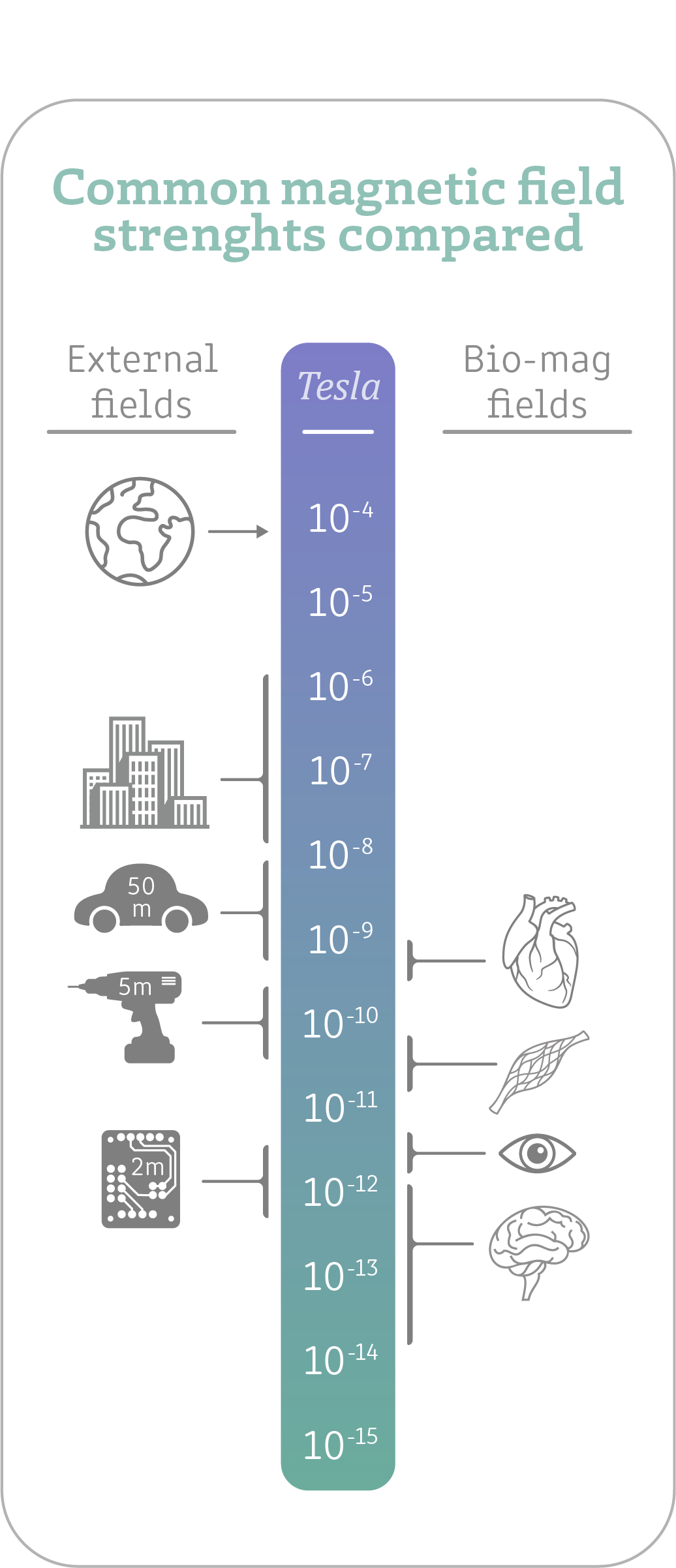
Image 3. Comparing the magnetic fields of different common sources.
Noninvasiveness
Along with its excellent temporal resolution the other main advantage of MEG is the non-invasiveness and patient comfort. The MEG scanner doesn’t need any preparation from the subject. The subject just removes all magnetic material from their person and then they can have a seat in the MEG scanner.
The subject is recorded either in a supine or seated position with the seated position being the norm. They are then positioned so that their head is up in the MEG helmet as close as possible to the sensors. This may first feel a bit uncomfortable but usually in a few moments the subject relaxes and habituates to the position.
Common MEG measurements are run by introducing a simple stimulus and running it at least 80-120 times over to average out any residual magnetic noise and uncorrelated neural background activity. At times, depending on the stimulus intervals and paradigm, the measurement might be a bit tiresome and boring. This might be a slight nuisance for the subjects.
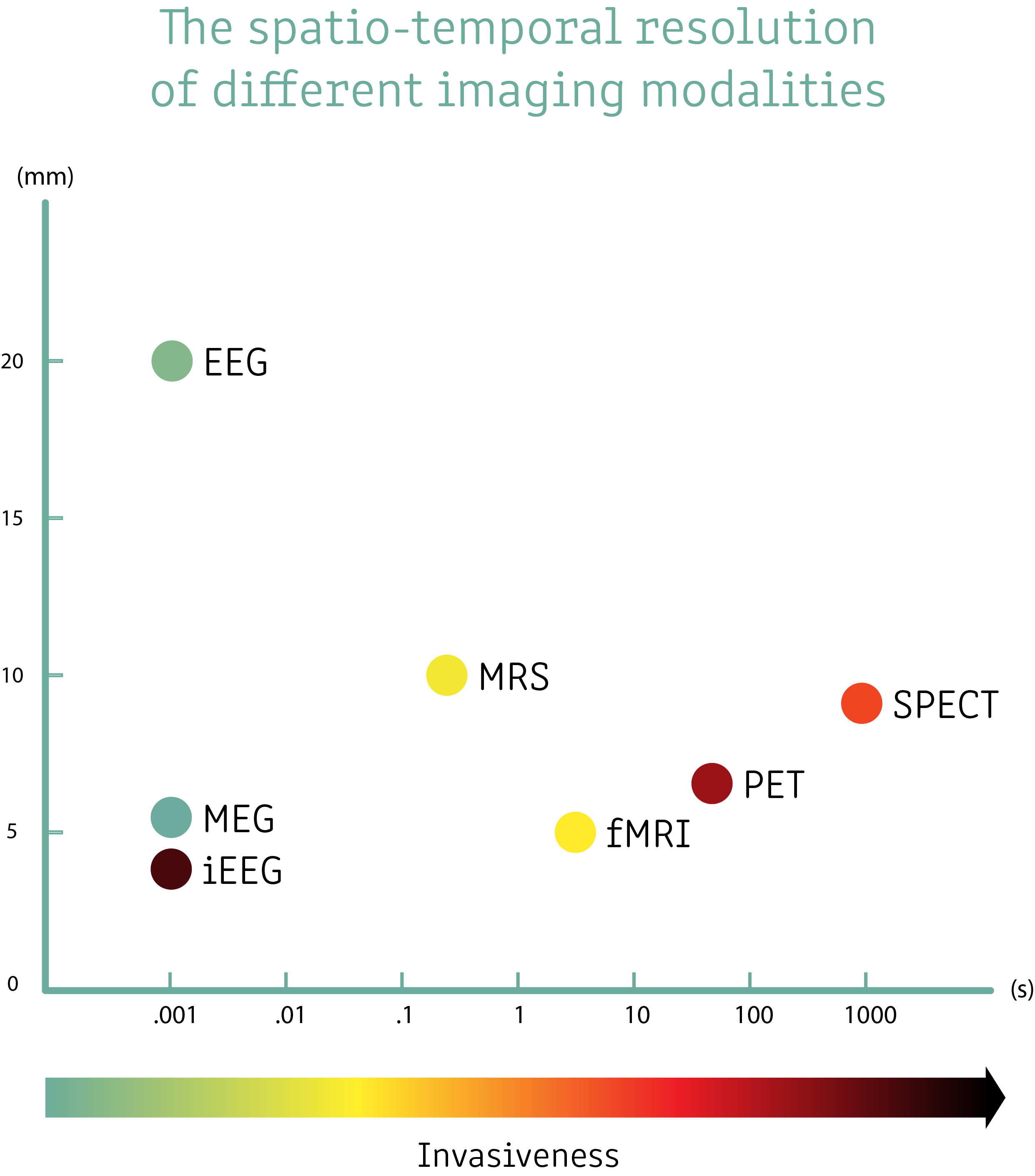
Image 4. Comparing the resolution of different modalities
Even though MEG measurements have its downsides it is still very non-invasive for the subject. Compared to MRI imaging it is much less restrictive. EEG needs usually 32-128 electrodes which have to have a good contact to the scalp. This makes EEG usually a bit more uncomfortable than MEG. Most other functional and anatomical imaging methods are generally seen as more invasive than MEG.

Image 5. A cartoon depicting a subject positioning themself under the MEG helmet. The measurement is very noninvasive and even somewhat comfortable. This is a clear advantage of the MEG measurement as opposed to other neuroimaging methods.
SQUID-sensors
How they work
SQUID stands for Superconducting Quantum Interference Device. The SQUIDs are in essence ultra-sensitive magnetic field sensors – magnetometers. The technology is based on superconducting loops with Josephson Junctions. With superconductors, an applied static magnetic field gives rise to a continuous shielding current on the surface of a superconductor, which in turn generates a magnetic field exactly cancelling the impinging external field, thus preventing the external field from entering the superconducting material. If the shielding current exceeds the critical current, a flux quantum may “slip” into the ring, elevating the flux to the next quantum flux state and lowering the shielding current accordingly.
Superconducting loops must be unbroken to maintain these characteristics, which makes it hard to measure the magnetic field applied to the loop. Thus, the Josephson Junctions are a solution to this problem. The ring is broken by a thin layer of electrical insulator that still allows for quantum tunneling of electron pairs. This allows an interference of the electron wave functions and this gives rise to a measurable quantity – flux-dependent resistance. With a constant bias current applied through the SQUID the average voltage over the SQUID is altered in a non-linear way. When the flux is changed through a SQUID it gives rise to a periodical voltage-flux function. Increasing the flux by one flux quantum does not change the SQUID output. In MEG the SQUIDs are coupled with a negative feedback system that generates an additional magnetic flux that opposes the actual flux. Since the feedback signal directly opposes the actual magnetic flux and is proportionate to the actual variations in magnetic flux, it can be used as the actual signal for the MEG acquisition.
Different types
The SQUIDs themselves are too small to be practical in MEG. Thus, the SQUID is usually coupled with a flux transformer. The idea is to couple the SQUID to a coil with a much larger area to increase sensitivity. To account for different needs in measuring brain signals, there are a few different kinds of flux transformers, varying the coil geometry and no. of coils. Typically, a flux transformer has a pick-up coil and a signal coil, with some having an additional compensation coil. The pick-up coil is located closest to the brain. The signal coil is on top of the SQUID loop. the optional compensation coil is further away from the brain, and it attenuates the magnetic fields originating from outside the head.
Magnetometers are simple sensors with a single pick-up coil and no compensation coil, coupled with the SQUID via the signal loop. Magnetometer is most sensitive to the magnetic component along the direction perpendicular to the surface of the pick-up coil. Magnetometers are very sensitive to brain signals, but they are also sensitive to far away magnetic sources.
To increase the acuity to brain signals as opposed to far away noise sources, a compensation coil can be added. This configuration is called a gradiometer. Older systems used axial gradiometers where the compensation coil was further away from the brain, but modern systems often use planar gradiometers with the coils being side by side. Orientating planar gradiometers 90° to each other gives us good spatial accuracy and the sensors complement each other.
In a modern thin film sensor, the different sensor types are stacked on top of each other in a single unit. The magnetometers are most sensitive to magnetic fields around the rim of the sensor, and thus couple strongest to neural sources next to the sensor. Planar gradiometers are most sensitive to distinctly oriented field vectors directly under the sensor.
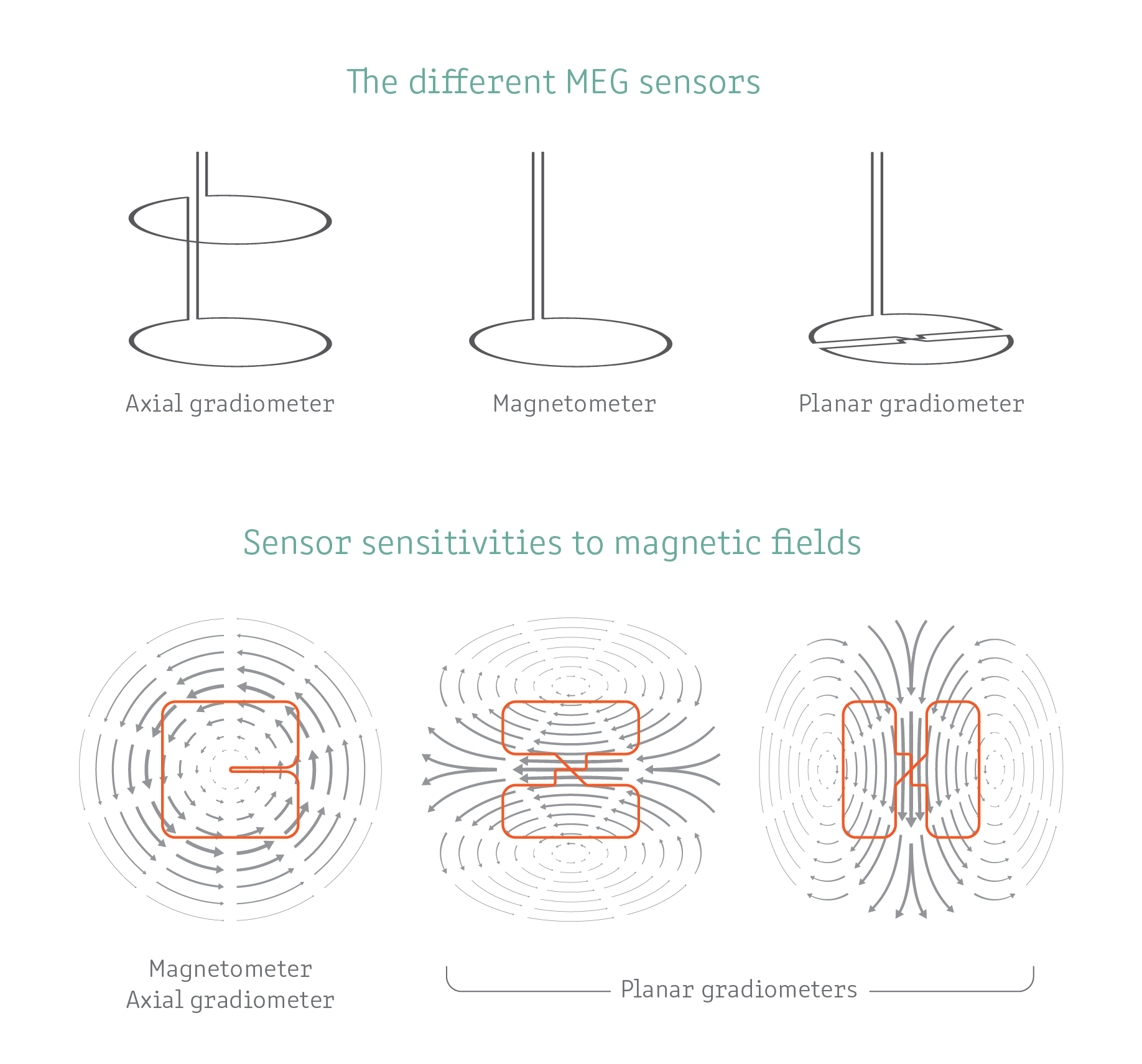
Image 6. Here we depict the geometry of the most common SQUID-sensor coils and also their relative sensitivities to magnetic fields. We can see that the magnegometer and axial gradiometer are most sensitive on the edge of the sensor and the axial gradiometers are most sensitive directly beneath the sensor.
Large sensor arrays
Modern MEG systems employ whole-head measurement arrays. There are a few manufacturers, but the one used in the Jyväskylä laboratories is the Elekta Neuromag TRIUX - 306 channel MEG. The channels are composed of 102 sensor units, each housing 3 different SQUID sensors. There are 2 planar gradiometers oriented perpendicular to each other and then a magnetometer in each sensor unit.
The sensors are placed evenly in a helmet shape. The idea is to capture signals from all over the cortex. The problem with making a helmet to fit ‘all’ different head shapes is that the magnetic signals attenuate rapidly with increasing distance. Thus, it would be best to get the subject’s head right next to the sensors. People with smaller heads will always have their cortex a bit further from the sensors in some direction.
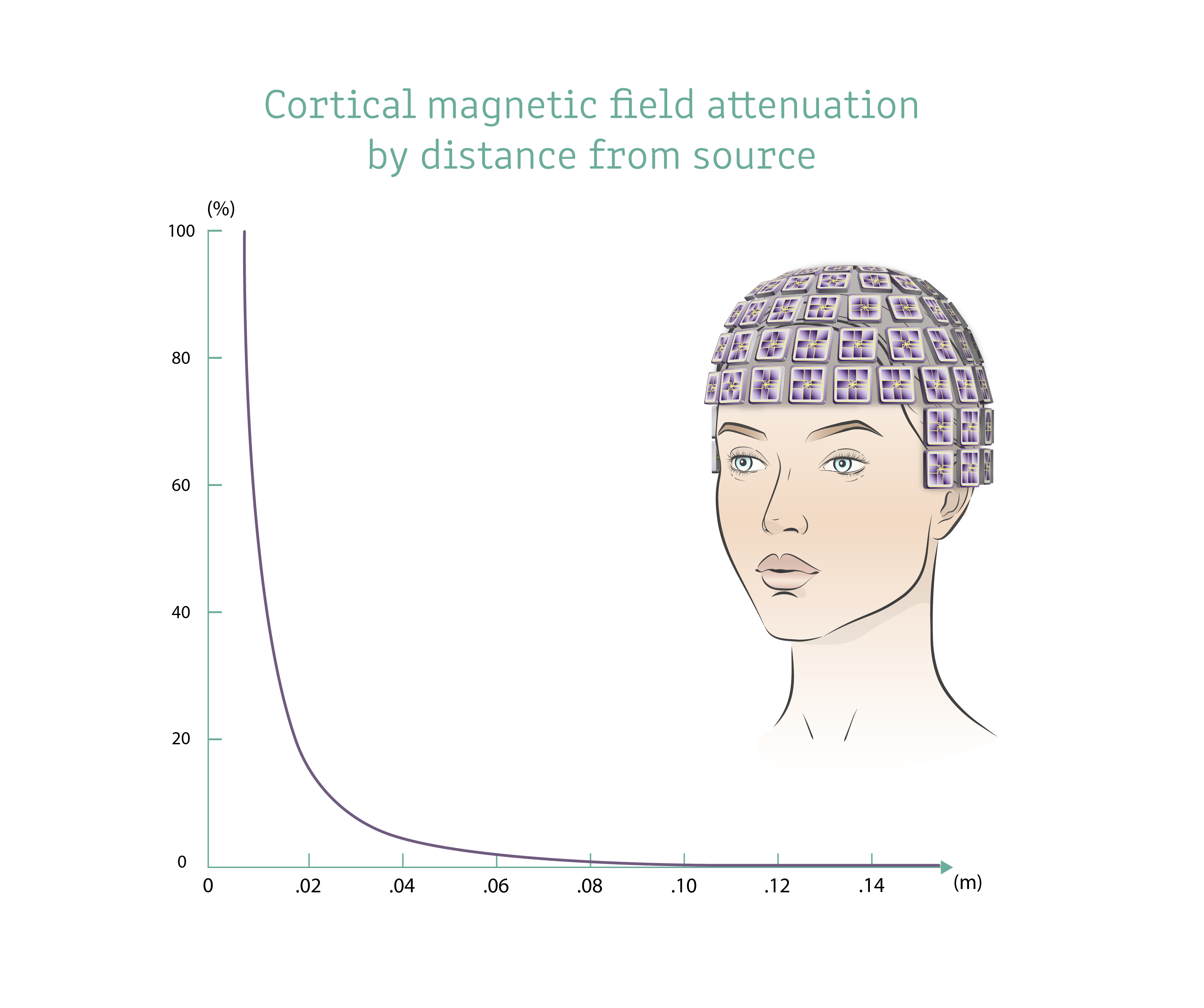
Image 7. The cortical magentic field strenght falls off rapidly with increasing distance. On the right, an artist rendering of a meg whole head system sensor geometry (modelled after the MEGIN 102 sensor array).
A typical measurement session
A typical measurement session starts with preparing the lab. The lab operator makes sure that all the necessary items are available for the measurement. The operator then connects all the required stimulation equipment and sets up the measurement environment according to the project’s specifications. For example, in a typical auditory evoked response measurement, the operator makes sure that the necessary audio equipment is ready and connected to the stimulus and measurement computers. It is also good to make sure that you are getting the stimuli triggers in the meg data.
When the subject arrives, it is good to give them a brief tour of the lab and answer any questions they might have. After the pleasantries, the subject is asked for a written consent. Then the subject is asked to remove any metallic/magnetic materials and they seated in the MEG scanner. This is done to ensure that the subject has no residual magnetic noise on their person.
The subject is then prepped by attaching necessary electrodes and head positioning coils. The subject’s head is registered in 3d with a stylus – this is called digitization. After the preparation the subject is once more seated in the shielded room in the MEG scanner. After all stimulation devices are set up and connected, the subject is hoisted into the MEG helmet as far as they can comfortably go. After this, the operator exits the shielded room and closes the door.
The MEG recording is started, and the stimulation protocol can begin. There is an intercom connecting the 2 rooms together and there is a video feed also. Typically, about 70-120 trials of a given stimulus are enough. After there is sufficient data from all the different trials in the protocol, the subject is assisted out of the MEG scanner and shielded room. In a longer measurement it is good to have pauses in between blocks.
After the subject leaves the data can be stored securely on servers. Then the lab is cleaned for the next measurement.
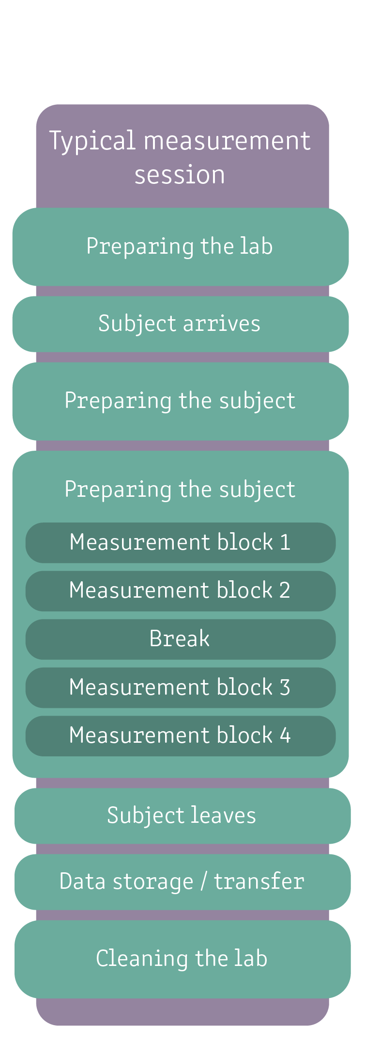
Image 8. An example of a typical measurement.
Possible participants
To avoid magnetic interactions – especially with metals – because they reduce the measurement accuracy, no metals should be worn during the measurements. Before entering the MEG room, the subject is inquired about any possible constraints which limit their participation in the study. The following are generally seen as contraindications for meg measurements:
- Any electronically controlled devices such as pacemakers, neuro-stimulators, insulin pumps or hearing devices.
- All metallic objects – can be discarded prior to measurements (e.g., keys, glasses, belts, …)
- Metallic implants also have the possibility to be ferro magnetic. The researcher needs to be informed about all known or possible implants prior to the measurement
- Tattoos (also permanent make-up) can contain color particles with metallic parts in them
- Cosmetics such as mascaras, rouge or even styling gel can contain metal. Please avoid using those on the day of measurement
- Glasses (contacts are fine), there are non-magnetic glasses available in the meg lab